

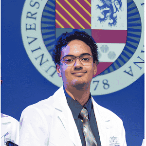
Speaker
Kevin Delzepich
- Creighton Medical School
- USA
Presentation
High-Resolution Imaging of Type I Truncus Arteriosus With Anomalous Left Coronary Artery Origin
- April 22-23, 2027
- Paris, France
Biography
Kevin Delzepich, third-year medical students at Creighton Medical School in Phoenix, Arizona. His current work examines cardiovascular imaging in congenital heart disease.
Abstract
Background:
Truncus arteriosus is a rare congenital cardiac defect in which coronary anatomical variations can complicate repair. Defining the coronary ostial position and proximal course is important for surgical planning and preserving myocardial perfusion.
Objectives:
We describe a posterior left coronary origin in type I truncus arteriosus and the contribution of computed tomography (CT) angiography to preoperative anatomical assessment.
Key Results/Findings:
A male infant born at 35 weeks presented with cyanosis and respiratory distress. Transesophageal Echocardiography demonstrated type I truncus arteriosus, a ventricular septal defect (VSD), and an atrial septal defect (ASD). Three days later, CT angiography with three-dimensional reconstruction confirmed the diagnosis and demonstrated an anatomical variation: the left coronary artery arose from the posterior (noncoronary) sinus rather than the left coronary sinus. The infant underwent truncus repair with ventricular septal defect patch closure, was transferred postoperatively to intensive care in stable condition, and was subsequently discharged with planned specialist follow-up.
Conclusion:
This case underscores the importance of high-resolution imaging techniques in diagnosing and managing congenital heart defects. While echocardiography serves as the initial imaging modality, clinicians should remain vigilant for less apparent defects. CT angiography with three-dimensional reconstruction can complement echocardiography by delineating coronary origins and their relationships to structures involved in repair. Identifying potentially vulnerable coronary anatomy may guide reconstruction and help avoid coronary injury, distortion, or compression, to preserve myocardial perfusion and reduce perioperative complications.
